

Ever wondered if a computer could spot a tumor before a doctor does? These days, smart systems help doctors study images from tests like X-rays and CT scans. Imagine a clever helper that sifts through hundreds of pictures in just minutes, catching tiny signs, like a small spot on the lung or a faint mark on the brain, that might be missed otherwise.
This means tests get done faster and images come out clearer, which eases the doctor’s workload and boosts care for every patient. AI in medical imaging isn’t just another tool; it’s changing the way we understand and take care of our health.
Transformative Effects of AI in Medical Imaging Diagnostics
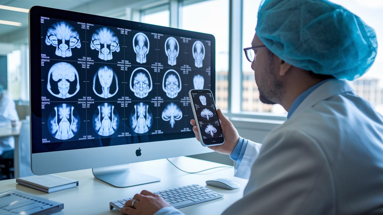
AI-powered tools are changing the way doctors study images in radiology, pathology, and cardiology. Using deep learning (a way for computers to learn patterns), smart machines quickly sift through loads of images. For example, these systems can pick out tiny lung nodules or brain tumors with more precision than older methods. Think of it as having a helpful assistant that looks over hundreds of pictures in just minutes, so doctors can work faster and reduce delays, especially after all the backlogs from the pandemic.
This new tech also means better handling of loads of data. With automation doing the routine work like outlining and labeling images, radiologists can focus on the tougher cases that really need their attention. Imagine getting a quick snapshot of a tricky chest scan, almost like a mini head-up before diving into a good story. That speed and clarity boost both the confidence of clinicians and the well-being of patients by smoothing out the treatment process.
AI in medical imaging isn’t only about spotting problems, it makes the whole process smoother. Advanced algorithms catch details that might slip past the human eye, cutting down the time needed for a full diagnosis. As more hospitals start using these smart tools every day, the future of healthcare diagnostics looks bright. We’re talking better outcomes for patients and less work for busy health professionals.
Core AI Techniques and Algorithms in Medical Imaging

Deep learning is a game-changer for making scans clearer. These clever tools use things like convolutional neural networks (CNNs) – a type of smart system that spots patterns super fast, even faster than we do! Picture a system that highlights the tiniest hints in a lung scan, turning a blurry picture into a clear one you can actually use. It cuts down on guessing and speeds up the process by cleaning up noisy images.
Traditional machine learning also plays a big part in helping with diagnoses. It works like a friendly helper that processes huge amounts of data step by step, much like sorting through a photo album to pick out the best shots. These methods break down images into parts, sort them, and even spot unusual areas. That way, doctors get a neat set of images they can trust, which means quicker and sharper treatment decisions.
The best part? Mixing deep learning with classic machine learning boosts both image quality and speed. Instead of painstakingly drawing lines by hand, AI can automatically highlight the important spots. This extra set of reliable eyes helps radiologists focus on the details that matter most, all while offering smart measurements to back up their calls. It really feels like having a tireless teammate by your side.
| Technique | Purpose |
|---|---|
| Convolutional Neural Networks | Pattern recognition |
| U-Net Architectures | Precise segmentation |
| Autoencoders | Noise reduction |
| Random Forest Models | Tissue classification |
| Support Vector Machines | Anomaly detection |
| Generative Adversarial Networks | Resolution enhancement |
Key Clinical Applications of AI-Driven Medical Imaging
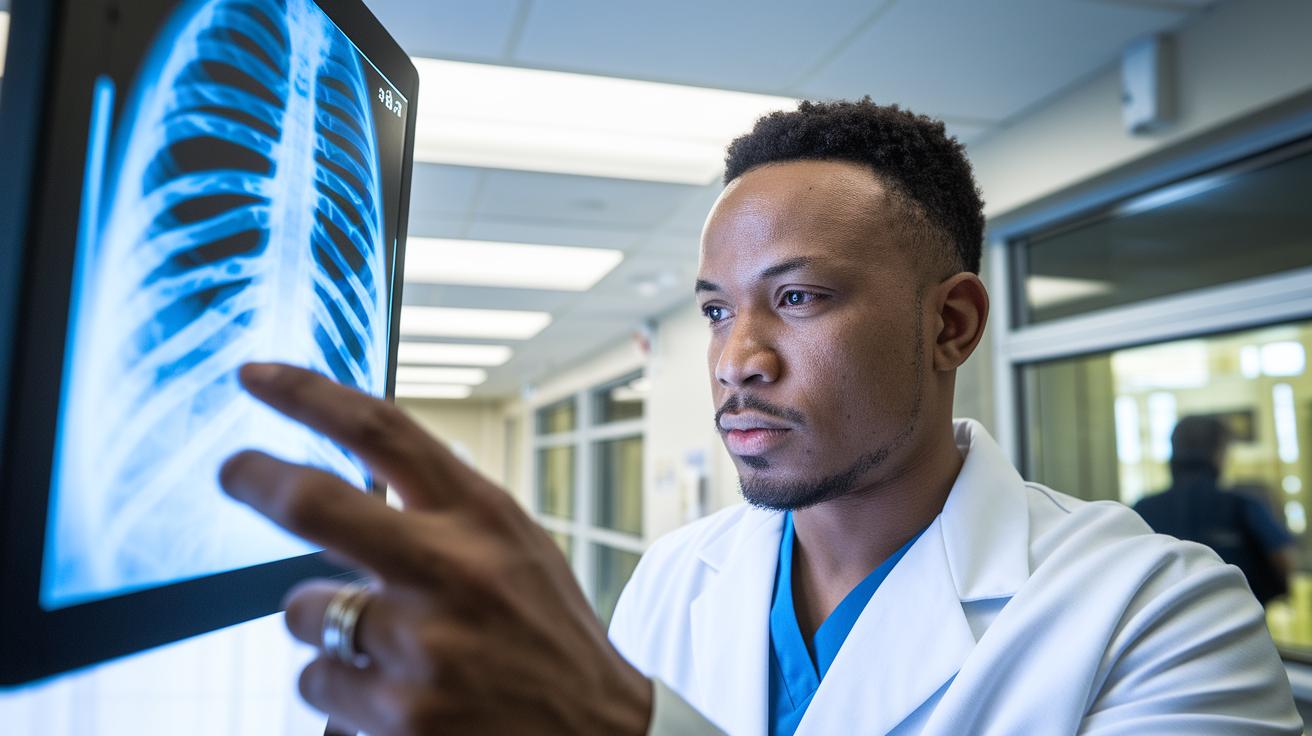
AI is changing the way we diagnose and treat by helping doctors look at images more clearly and make faster decisions. In radiology, smart systems check chest X-rays for heart issues and scan CT images to quickly spot fractures. Imagine a busy emergency room where every moment matters, when an AI flags a possible fracture, doctors can jump in immediately. One radiologist even said it was like the tool softly hinted, “Hey, look here,” catching something they might have missed.
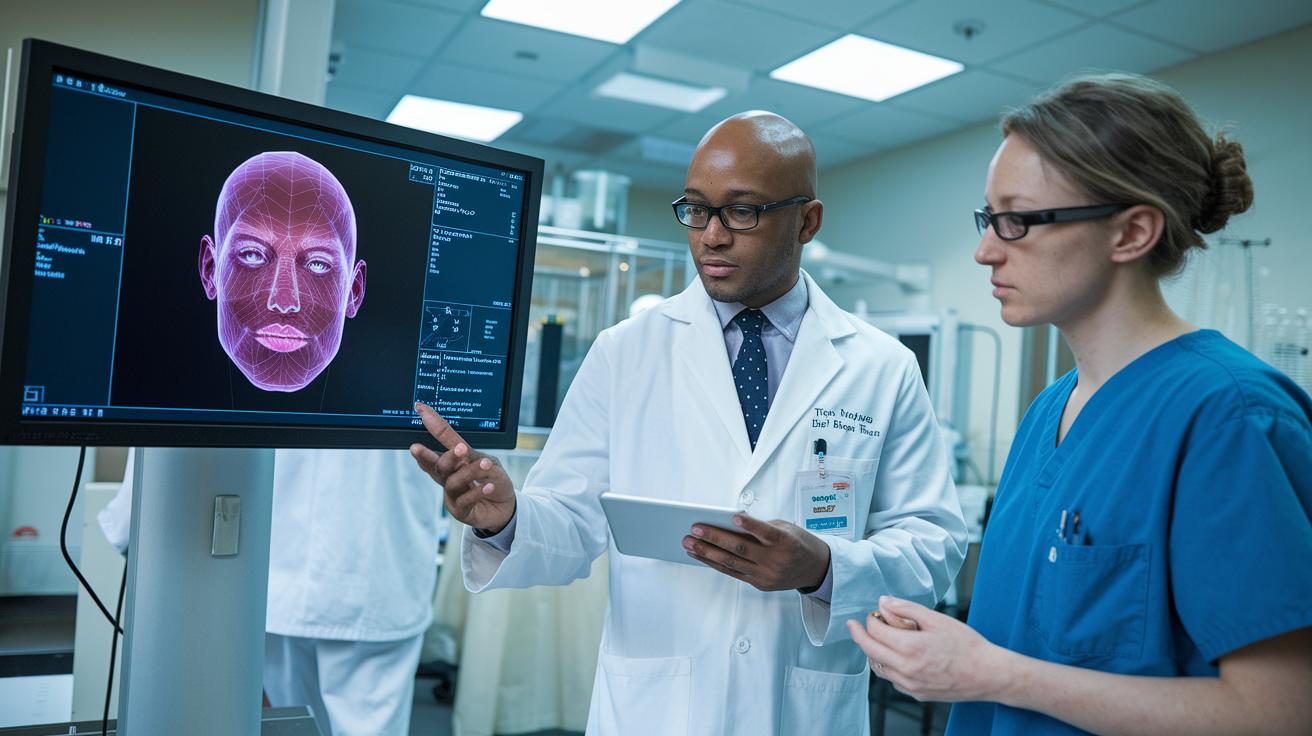
In pathology, AI looks over histology slides to pick up tiny details in cells. It can quickly go through many images and point out areas that need a closer look. For example, while examining a slide from someone suspected of having cancer, the AI might notice a group of unusual cells that deserve extra attention. This extra check helps build trust in the final diagnosis.
Cardiology is another area where AI is a big help. Advanced systems study echocardiograms to spot valve issues and properly measure heart function. Picture a cardiologist with a pile of scans, AI makes it easier by giving precise numbers and details about the heart, so treatment can be tailored to each patient. And that's not all. AI tools are also used to assess bone injuries, guide careful procedures, and keep track of long-term conditions with clear data.
Other useful applications include:
| Application | Description |
|---|---|
| Fracture Risk Assessment | Automatically analyzing scans to identify risk areas |
| Minimally Invasive Guidance | Providing enhanced image overlays to guide procedures |
| Chronic Condition Tracking | Continuous evaluation with data-driven insights |
By adding these smart tools to their routine, doctors get clear, structured info that helps them make quick, informed choices. This not only sharpens the diagnosis but also makes healthcare safer and more efficient, ensuring every patient gets the benefit of improved accuracy and rapid care.
Challenges and Regulatory Considerations for AI in Medical Imaging

One big challenge is having access to diverse, high-quality imaging data. When data comes from just one type of source, the AI might work well for some people but not for others. Imagine an AI tool that catches big issues in scans but misses smaller signs in images from groups that aren’t well represented. This gap raises big ethical questions and shows why we need to gather varied data to build fair AI systems.
Another important point is that the AI needs to be explainable. Health professionals need to understand how the algorithm reaches its decisions. It’s like following a map where every turn is clearly marked. Without clear, understandable steps, trust can start to slip. When an AI system explains itself, doctors feel more confident in their diagnosis and patients get a clearer picture of their care.
Regulatory rules from bodies like the FDA and CE are still trying to keep up with new tech advances. Sometimes, outdated rules slow down the safe rollout of smart diagnostic tools. For example, after the pandemic, imaging services were overwhelmed. This shows why updated standards are so important to confirm that AI tools are safe for use in clinics. It means we need to revise how we validate and monitor these systems to match the fast pace of technology and growing demands.
People in the medical community are actively discussing new regulations that build trust, protect patient safety, and make sure that smart tools work fairly across everyone.
Workflow Integration and Automation in AI-Powered Imaging

AI is reshaping everyday work by handling tasks like image segmentation and annotation. Picture software that spots lesions on a CT scan and drafts a basic report in seconds. This leaves radiologists free to dive into the details that really matter. Plus, automated systems create clear, consistent data for systems like PACS (the archives where images are stored), so every scan comes with standardized info.
Getting these smart tools up and running takes a few careful steps. First, departments run pilot tests to see how the new tech fits with their current routines. Next, staff training sessions help everyone feel at ease using the system. Then, IT integration links the new tools with existing radiology and picture archiving systems. Think of these steps as key checkpoints to make sure the transition runs smoothly:
- Pilot testing to check real-world performance
- Staff training to build confidence and expertise
- IT integration to ensure data flows easily with existing platforms
- Setting up standard reporting templates for consistency
- Ongoing monitoring to fine-tune the workflow
By automating routine tasks, AI-powered tools streamline clinical work. Radiology departments enjoy more accurate reports and quicker turnaround times, easing the pressure of diagnostic backlogs. And with fewer manual errors, clinicians can focus on the critical cases, ultimately boosting patient care in our increasingly digital world.
Future Research Trends and Innovations in AI-Driven Imaging
Research in AI imaging is truly reshaping the way we think about medical diagnostics. Academic studies and industry trials are teaming up to create smarter systems that learn from a vast mix of real-world data. Researchers are blending different AI techniques with tools that can even predict how a disease might progress. Imagine a system that can forecast wound healing or alert doctors to the risk of diabetic foot issues early on, allowing for quicker, potentially life-saving actions. All of this is backed by an ever-growing collection of studies that show how raw data can be turned into clear, actionable insights for healthcare professionals.
There’s real excitement among health experts about early tests that combine imaging signals with clinical information. These aren’t just theoretical ideas, this trend is paving the way for automated imaging that boosts both accuracy and speed when diagnosing patients. Recent studies suggest that gathering data from various imaging methods leads to a more complete picture of complex conditions. It’s almost like having a group of experts pooling their knowledge together, with advanced computer vision techniques as the common thread.
RSNA Resources for Advancing Imaging AI
RSNA is helping drive this innovation by offering a range of educational platforms. Their programs include opportunities for grant writing, detailed guides for research development, and even an Imaging AI Certificate. They also provide curated case libraries where trained models are sorted by specialty, giving both researchers and clinicians a handy toolkit to rely on.
Emerging Innovations in Computational Diagnostics
Investments in computational diagnostics are sparking fresh interest in bringing different types of data together. Early research highlights new imaging markers and points to emerging markets that are ready for breakthrough innovations. This momentum is pushing AI research towards more accurate and efficient imaging solutions.
Final Words
In the action, we explored how artificial intelligence in medical imaging reshapes diagnostics. The blog covered key AI techniques that sharpen imagery and improve report speed. We reviewed its clinical uses, from spotting lung nodules to flagging tissue issues, and discussed challenges like data equity and regulatory updates. We also touched on integrating AI into daily workflows to free up time for radiologists. It’s clear that these smart advances offer real benefits, setting a positive tone for easier, more precise healthcare management.